Science · Built from a recipe
Super-Res Microscope
C4 on the minor comb. Its drum rings at 252.7 Hz.
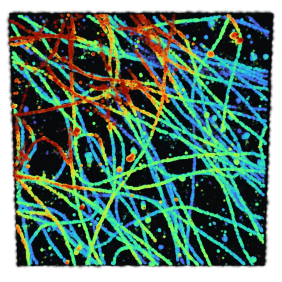
Ordinary light microscopy struggles to separate nearby objects about 200 to 300 nanometers apart.
- Shelf
- Science
- Kind
- Built from a recipe
- Your own
- Your own localizations
- Sound
- Yes
- Licenses
- CC BY 4.0
Its note
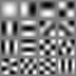
Splashery stretches a drumhead over this Splash's outline, twelve inches across, and finds the ways it can ring. Its lowest is 252.7 Hz, 60 cents flat of C4, the nearest tine on the comb. The next fifteen are its overtones, and each one draws its own figure: sand gathers on the lines that stay still.
- 1253 Hz
- 2399 Hz
- 3400 Hz
- 4506 Hz
- 5563 Hz
- 6566 Hz
- 7644 Hz
- 8644 Hz
- 9733 Hz
- 10738 Hz
- 11757 Hz
- 12798 Hz
- 13800 Hz
- 14890 Hz
- 15894 Hz
- 16906 Hz
How to play
Pinch or scroll to zoom down to single molecules. Tap to see a thin slice at one depth.
Its controls
| Control | Kind | Range | Start |
|---|---|---|---|
| Show a slice at the depth you tap | Tap | ||
| A slice at one depth | Switch | Off | |
| Data | List | 7 choices | Microtubules and clathrin |
| Color by | List | 3 choices | Depth (z) |
| Depth scale | List | 2 choices | True (1×) |
| Precision | List | 3 choices | All localizations |
About it
Ordinary light microscopy struggles to separate nearby objects about 200 to 300 nanometers apart. Single-molecule localization microscopy finds the centers of isolated fluorescent blinks more precisely, then builds a picture from many positions. Localization precision is not the same as the final image's resolution, and several blinks may come from one molecule. Here each blink is drawn as a Gaussian whose width represents its reported uncertainty.
Choose microtubules and clathrin in a 12 µm cell region, a whole nucleus in 3D, or open a .smlm file or a CSV with positions and uncertainties in nanometers. Color by depth, time frame or channel. Depth scale shows true coordinates or stretches depth fourfold. Precision keeps all localizations or only those better than 5 or 3 nm. Pinch or scroll to zoom; tap for a 200 nm slice at that depth, then tap to restore all depths. Files stay on your device. This is a viewer, not a measurement tool.
Details
- Splats
- Opens at 200,000 at Mid Detail, 84,000 at Low, and up to 200,000 at Max
- Your own files
- .smlm, .csv, .txt, .zip
Sources and credits
| Work | By | License |
|---|---|---|
| Microtubules and clathrin in a Cos cell (ShareLoc.XYZ, 10.5281/zenodo.5507427) | Christophe Leterrier (Aix Marseille Université, CNRS, NeuroCyto), on ShareLoc.XYZ; a 12 µm square cut from the record (a subset) | CC BY 4.0 |
| Zola-3D NUP full nucleus (ShareLoc.XYZ, 10.5281/zenodo.7233696) | Andrey Aristov (Institut Pasteur), uploaded by Benoit Lelandais, on ShareLoc.XYZ; a random half of the localizations (a subset) | CC BY 4.0 |
| Xenopus laevis nuclear pore complex stained with WGA-ATTO520 (ShareLoc.XYZ, 10.5281/zenodo.7182237) | Anna Löschberger, on ShareLoc.XYZ; an 8 µm square cut from the record (a subset) | CC BY 4.0 |
| Actin with PhalloidinAF647 in COS7 (ShareLoc.XYZ, 10.5281/zenodo.5510661) | Sarah Aufmkolk, on ShareLoc.XYZ; a 10 µm square of one cell, 174,903 of its localizations (a subset) | CC BY 4.0 |
| Mitochondrial protein TOM22 in COS7 cells (ShareLoc.XYZ, 10.5281/zenodo.5512636) | Wei Ouyang, on ShareLoc.XYZ; a 15 µm square of one field, 219,846 of its localizations (a subset) | CC BY 4.0 |
| ZOLA-3D microtubules (ShareLoc.XYZ, 10.5281/zenodo.6861446) | Andrey Aristov, Benoit Lelandais and Christophe Zimmer (Institut Pasteur), on ShareLoc.XYZ; a 12 µm square, 159,785 of its localizations (a subset) | CC BY 4.0 |